ELECTRON MICROSCOPY [home] [topic page]
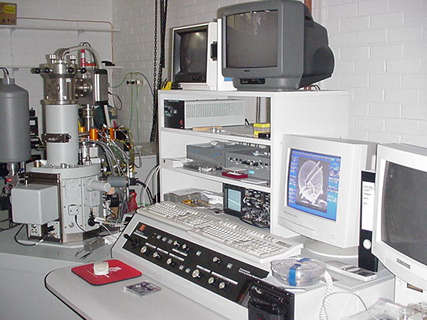
University of Tasmania's Central Science Laboratory scanning electron microscope.
Using electrons instead of light to view small objects has many advantages.
All microscopes work by
Optical microscopes use glasses to achieve the effect of a larger image, electron microscopes use magnetic and electrostatic fields.
Visible light is limited. The ability to see small detail is governed by an effect called "diffraction". As light passes around or is reflected by a small object, it "smears" a little. The amount of smearing is governed by the size of the thing studied, the object, and the wavelength of the light. For this reason, optical microscopes cannot, even in principle, see details much smaller than the wavelength of light itself, about 0.4 micrometres.
Electrons, however are a different kettle of fish. They, through quantum mechanics, can be allocated a wavelength which depends directly on their speed from the source. The faster they travel, the smaller their wavelength so the smaller their diffraction limitations.
From the de Broglie equation
h / l = momentum, l = wavelength , h = Planck's Constant
it follows that there wavelength is given by
l = h / momentum.
Faster electrons have greater momentum thus l is smaller.
Simply accelerating the electrons at a higher speed to begin with ensures greater detail.
Further, electrons are charged and have mass. They therefore can be manipulated differently to create different types of images for study.
The microscope above is a "scanning electron microscope" ( SEM ) . Electrons, instead generally washing over the object, as would be the case normally, are traced back and forth in a narrow beam over the surface of the object.
After the electrons strike the target, many signal radiations are produced. The main ones used are backscattered and secondary electrons. Backscattered are the high energy incident electrons which have "bounced" off atoms. Secondary electrons are electrons knocked out of atomic or molecular configurations.
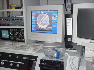
These electrons are detected and their number represents the "brightness" as the scan proceeds. The monitor display scan rate is locked to the scan rate of the object, the backscatter current is the brightness of the display.
As the microscope works, not by transmitting electrons through the object, but by reflection processes, the object of study must be made into a conductor, often by gold plating it. The whole assembly is placed in a vacuum so the electrons can easily traverse the microscope.
Typical resolution of these machines is 10nm, about 100 atoms. Electron gun PD may be about 2kV.
Excellent web sites for studying the SEM are at
http://www.mse.iastate.edu/microscopy/home.html
