Nuclear Magnetic Resonance NMR [home] [topic page]
A technique used to determine the structures of molecules.
This technology is very recent and relies on the detailed knowledge of the construction of nuclei of various atoms. In turn it relies on quantum mechanics and quantum electrodynamics. NMR was discovered in 1947 but these machines have only been possible in the last 40 years. They have come out of nuclear research facilities into medicine and chemical analysis as cheap computing has become available.
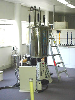
University of Tasmania's Central Science Laboratory NMR machine. This runs at 9.4T. The large space around it is because of stray B fields which can corrupt computers, pacemakers etc.!
NMR relies on
- a very strong magnetic field - often using superconductors (ref 1,)- typically of the order order of 1~10 tesla.
- nuclei of the correct "spin" value
- applied radio waves
- detectors of the resulting emission
- computers and programs capable of translating the emissions into understandable data.
Most systems are set up to detect protons or C- 13 nuclei.
So - what is it all about?
Protons and neutrons "spin" and behave as small magnets to a tiny extent!
Key point; moving charge creates magnetic fields, spinning is moving. Why do neutrons then behave as a magnet? Neutrons are made of 3 quarks and so the charge associated with the neutron, while totaling zero, is not symmetric. One side can be thought of as slightly positive while the other slightly negative. Spinning this gives the magnetic field.
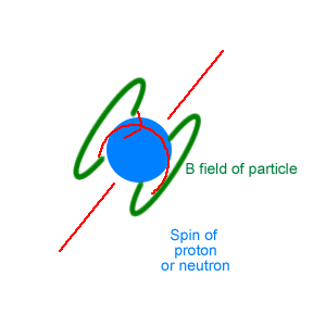
( PS - "spin" is not a very good word for what is happening here - it is strictly a "property" of the subatomic particles, a quantum number.)
Protons and neutrons each have a spin of size "1/2". To get the total spin of a nucleus, we must add the spins of all the nuclear members of an isotope vectorially. This in turn gives the nuclear magnetic moment of the nucleus.
If the total spin of a nucleus is 0 then NMR cannot detect the nucleus. O-16 and C-12 are examples of spin 0 so cannot be detected. This is unfortunate as these are very common atoms.
Spin of 1 includes H-2 and N-14.
Spin of 1/2 include H-1, the proton and C-13. These are the most commonly used nuclei in NMR.
So far so good!!!
We immerse the sample in the very strong magnetic field and this aligns the nuclei which have spin, like a compass needle aligning with the Earth's field. It is a little more complex of course as we are playing with truly tiny objects so quantum effects are everywhere. As a result, it turns out that the alignment takes a number of patterns depending on the total spin. Each alignment has a different energy.
We now "rattle" the nucleus with a pulsed radio wave. Pulses can be shown to cover a huge range of frequencies ( through Fourier analysis ). The correct frequency is applied during this pulse and the nucleus jumps from one alignment ( read energy ) to another alignment. This is a "resonance" similar to when a person pushes a swing a the same rate as the swing's period - the swing gets higher.
The Central Laboratory machine in the photo above pulses between 400-900 MHz with a pulse duration of 10ms. The power rating is 40 W.
Once the "resonance" occurs, the nucleus reradiates a radio wave photon with an energy equal to the difference between the states as it reverts to the lower energy alignment. The photon has a frequency related to the energy through E = hf where h = Planck's Constant and f = frequency. The more nuclei, the stronger the reradiation.
Because these photons are in the radio frequencies, an antenna can be used, a loop of wire, to detect them. ( This is not very different from the antenna of a car radio.) The photons become an electrical signal into the computer.
EFFECT OF ELECTRONS AND BONDS
The "resonance" is slightly modified when other atoms are near due to all the electrons. These include the bonding electrons. The emitted photons are then of slightly different frequencies than the naked atom's values. Electrons are magnetic because they have charge and spin. By looking at these photons from the unknown molecules and comparing them to known molecules, the structural bonds can be deduced. Further the relative strengths of the signals lead to the relative number both of bonds and of atoms. With this information, the molecular structure can be elicited.
MAGNETIC RESONANCE IMAGING is a closely related technology in which the reradiated signals from the nuclei are analysed spatially with powerful tomography software. This gives the position of the nuclei allowing pictures of concentrations of the atoms of interest to be created. It is a process widely used in medicine to create pictures in slices thence 3D constructions of sections of bodies.
The University of Tasmania's Central Science Laboratory's machine is fully capable of carrying this out on solids such as cuttings of plants.
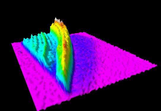
A 3-D visualization of the proton densities in a sandalwood cutting from water and oils.
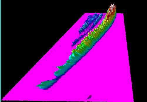
Oil densities in the same sample of sandalwood using the chemical shift effect (for oil only). It shows that most of the mobile protons in the wood are from the Sandalwood oil. It also shows that there is seasonal variation (concurrent with growth rings) and a tendency for the oil to be at highest concentration adjacent to the sapwood (the low level, dark blue region in the 1st image).
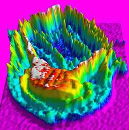
This is a straight proton density visualisation of a gall in a juvenile blue gum stem. ( Courtesy University of Tasmania Central Science Laboratory, Dr Evan Peacock)
( Unusual NMR - If a big enough sample exists, a strong B field is not needed. At the South Pole, NMR is being used directly on core ice samples using the vertical B field of the Earth! Large amounts are available so the reemitted radiation is strong enough to give useful signals.)
